01 / 06 SCIENCE
Lederman Lab
Thin-film growth & nanoscale surface characterization.
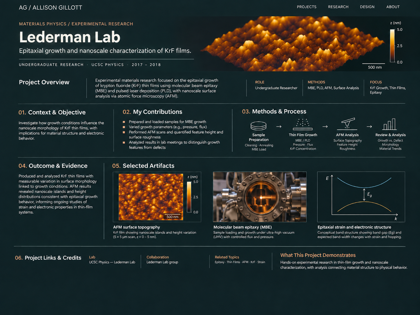
Project overview. At UC Santa Cruz’s Lederman Lab, I contributed to experimental thin-film research through sample preparation, molecular beam epitaxy (MBE) growth work, and atomic force microscopy (AFM). My role connected the practical demands of deposition with quantitative measurements of nanoscale surface morphology.
Understanding what thin-film growth leaves behind.
Thin-film experiments depend on controlled preparation and careful characterization. The surface that forms after deposition can contain meaningful growth features alongside contamination, defects, or imaging artifacts. Our work involved preparing samples and examining their topography to help evaluate film morphology within a broader materials-research effort.
From the growth chamber to the height map.
I worked primarily with fellow undergraduate researchers, with guidance from graduate mentor Becker and review by the wider lab group.
- 01
Preparation & growth
Cleaned and thermally prepared substrates, loaded samples for MBE, and participated in growth work that involved chamber-condition adjustments.
- 02
Surface characterization
Acquired AFM scans and measured nanoscale feature heights and surface roughness.
- 03
Collaborative interpretation
Reviewed height maps and possible anomalies with researchers; also assisted with PLD equipment setup as a separate, secondary activity.
Prepare. Grow. Measure. Question.
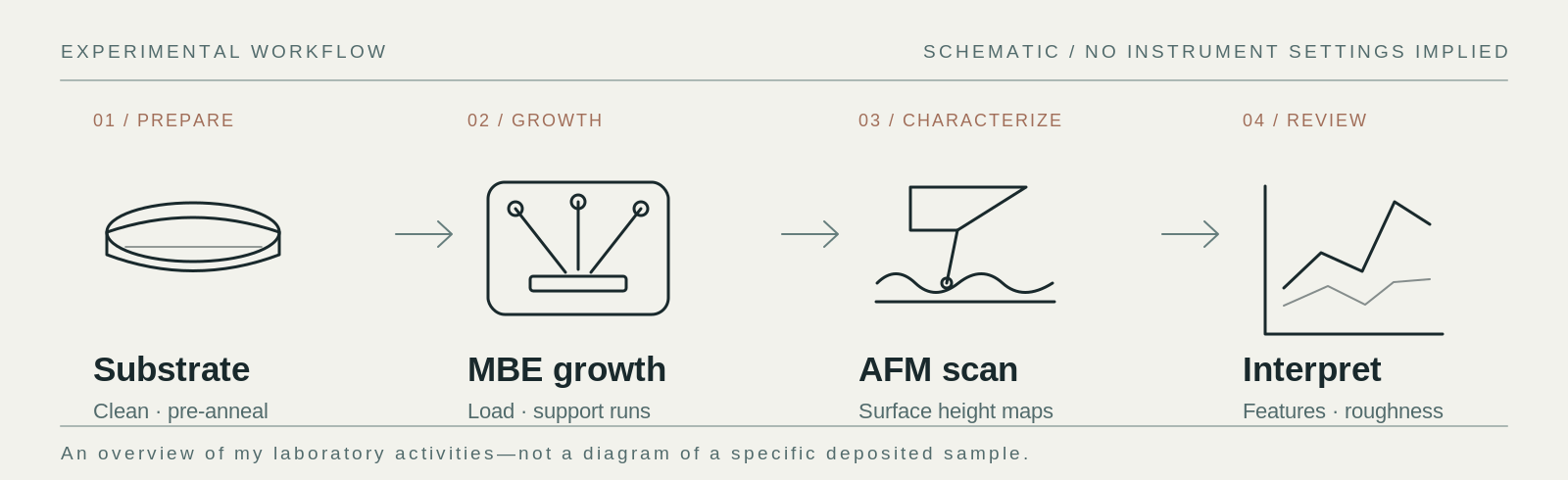
MBE work began with substrate preparation and loading. After growth, AFM translated the resulting surface into a height map—useful for comparing morphological features, but not a complete description of the material’s structure.
Prepare & deposit
Cleaning, pre-growth annealing, sample loading, and hands-on MBE work.
Scan & quantify
AFM imaging, feature-height measurement, and surface-roughness assessment.
Review & interpret
Compare observed hills or islands with possible defects, contamination, and scan artifacts.
What the surface reveals—and what it cannot.
My work contributed to the lab’s preparation and characterization workflow, producing AFM surface-height images and quantitative observations used in group discussions. The central lesson was one of measurement: a height map describes topography, while claims about composition, lattice strain, or electrical behavior require other evidence.
Substrate preparation, MBE involvement, AFM scanning, feature-height and roughness measurement, and collaborative review.
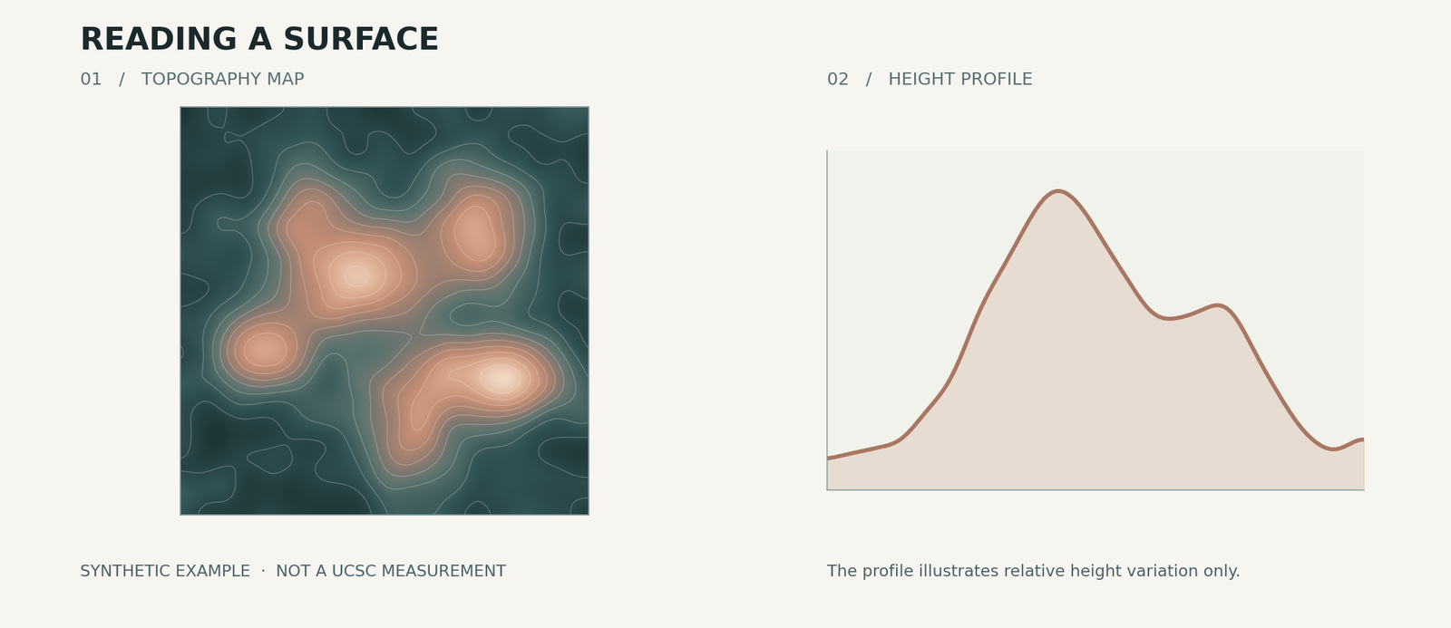
Original scans are not included in this V1. All visuals below are explanatory reconstructions—not measurements or findings attributed to me.
The method, visualized.
Original diagrams reconstruct the work’s sequence and the type of information AFM provides. They are not substitutes for the lab’s experimental data.
Lab context & references.
Hands-on experimental preparation and nanoscale characterization, with attention to the boundary between an observation and the interpretation it can support.